
AI in Medical Imaging: How Machines Analyze X-Rays and Scans
How AI Is Transforming Medical Imaging and Radiology
Medical imaging helps doctors see inside the human body without surgery. X-rays reveal broken bones. CT scans show detailed cross-sections of organs. MRI scans provide clear views of soft tissues. Ultrasound uses sound waves to create images of internal structures.
However, reviewing these images takes time, training, and careful attention. Doctors may need to examine hundreds of images to find a small abnormality. This is where AI in Medical Imaging can make a real difference.
Artificial intelligence allows computer systems to analyze medical images and identify patterns that may be difficult to notice at first glance. These systems can highlight suspicious areas, measure structures, compare image features, and help doctors prioritize urgent cases.
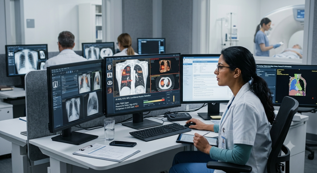
For example, an AI system may flag a possible lung nodule on a CT scan or identify signs of a fracture on an X-ray. A radiologist can then review the highlighted area and decide what it means for the patient.
AI doesn’t simply replace human judgment. Instead, it provides additional information that can support clinical decisions.
The technology is already being explored and used across different areas of healthcare. As medical imaging technology advances, AI-powered tools may help hospitals manage growing workloads, support earlier disease detection, and improve the consistency of image interpretation.
In this guide, we’ll explain how AI analyzes X-rays and scans, which technologies make it possible, where it is used, and what challenges healthcare providers must consider.
1. What Is AI in Medical Imaging?
AI in medical imaging refers to the use of computer systems that analyze medical images and identify useful patterns or features. These images may come from X-rays, CT scans, MRI machines, ultrasound equipment, and other diagnostic imaging systems.
Traditional medical image interpretation depends mainly on a trained professional examining the images. AI adds a computer-based layer of analysis that can help detect abnormalities, measure changes, or organize findings.
Understanding Artificial Intelligence in Medical Imaging
Artificial intelligence is a broad field of computer science. It includes systems that perform tasks usually associated with human intelligence, such as recognizing patterns and making predictions.
In radiology, AI systems are trained using medical images and, in many cases, expert-provided labels or clinical outcomes.
For example, developers may train a system using thousands of chest X-rays labeled by medical experts. The system learns to associate certain image patterns with findings such as lung opacities or pleural effusions.
After training and testing, the system can examine new images and estimate whether similar patterns are present.
These predictions are not the same as a confirmed diagnosis. Image quality, patient history, symptoms, and other clinical information still matter.
How AI Medical Imaging Differs From Traditional Analysis
Traditional image analysis relies on the knowledge and experience of radiologists. AI medical image analysis uses computational models to examine image features at scale.
The two approaches can complement each other.
| Feature | Traditional analysis | AI-assisted analysis |
| Image review | Performed by a medical professional | Supported by automated analysis |
| Pattern detection | Relies on clinical expertise | Uses patterns learned from data |
| Image measurement | Often performed manually or with software | Can automate selected measurements |
| Workflow | Depends on local processes | May prioritize or route selected cases |
| Final clinical judgment | Made by qualified clinicians | Requires appropriate human oversight |
AI can process many images quickly, but speed alone doesn’t guarantee a correct result. Its usefulness depends on the quality of the model, the images, and the clinical setting.
Why Medical Imaging Needs AI
Healthcare systems produce large volumes of imaging data. Radiology teams must review these studies while balancing urgent cases, routine examinations, reporting deadlines, and communication with other clinicians.
AI can help reduce repetitive tasks. It may also identify cases that need faster review.
For instance, a system trained to detect possible brain bleeding on CT scans could flag a suspicious study for urgent assessment. The radiologist would still review the scan and confirm the finding.
This is one reason AI-powered medical imaging is becoming an important area of healthcare innovation.
2. How AI Analyzes X-Rays and Scans: A Step-by-Step Guide
Understanding how machines analyze X-rays and scans makes the technology easier to understand. Although individual systems differ, most follow a similar process.
Step 1: Collecting Medical Images
The process starts when a patient undergoes an imaging examination.
An X-ray machine, CT scanner, MRI scanner, or ultrasound device creates images of the body. These images are usually stored in a digital format that medical imaging systems can process.
Many hospitals use the Digital Imaging and Communications in Medicine (DICOM) standard to store and exchange medical images.
The AI system receives an image or imaging study through an approved clinical workflow.
Image quality matters at this stage. Motion, poor positioning, noise, and incomplete scans can affect the analysis.
Step 2: Preparing the Images
Before analysis, software may check the image format, dimensions, and quality.
Depending on the system, preprocessing may include:
- Adjusting image intensity or contrast.
- Resizing images to match model requirements.
- Separating relevant image regions.
- Checking for missing or corrupted data.
- Identifying the correct body part or imaging series.
These steps help prepare the data for analysis. However, excessive or inappropriate image processing can also remove useful clinical details.
Step 3: Identifying Patterns With AI
The prepared images are passed to a trained model.
Deep learning models can learn patterns from large collections of examples. These patterns may include shapes, edges, textures, density changes, and relationships between different image regions.
For example, an AI model trained to detect lung nodules may learn to recognize small, rounded structures that require further examination.
The model doesn’t understand an image in exactly the same way a human does. Instead, it calculates patterns and relationships based on what it learned during training.
This process is often called automated image analysis.
Step 4: Detecting Possible Abnormalities
After analyzing the image, the system may generate an output.
Depending on its design, this output could include:
- A highlighted region on an image.
- A probability score for a particular finding.
- An estimated size or volume.
- A classification of possible abnormalities.
- An alert that a study needs urgent review.
For example, an AI tool used for CT scan analysis may highlight a possible lung nodule and estimate its dimensions.
The result is a prompt for further assessment, not automatic proof that a disease exists.
Step 5: Presenting Results to Radiologists
The system may display its findings within a radiology workstation or another clinical application.
A radiologist can compare the AI output with the original images, check whether the finding makes clinical sense, and consider the patient’s history.
If the AI misses an abnormality or highlights a normal structure, the radiologist must be able to recognize that possibility.
Step 6: Supporting Diagnosis and Reporting
Once the radiologist reviews the study, the findings can be included in the clinical report when appropriate.
Some systems can also assist with measurements, image comparisons, or draft report preparation.
However, AI-generated reports must be checked for accuracy. Incorrect measurements, missing findings, or misleading text can affect patient care if they go unnoticed.
Step 7: Monitoring Performance Over Time
AI systems need ongoing quality checks after deployment.
Hospitals should monitor whether the system works well with their scanners, patient populations, image protocols, and clinical workflows.
Changes in equipment or patient demographics can affect performance. Regular evaluation helps identify these problems before they cause wider issues.
In simple terms: AI receives an image, processes its data, looks for learned patterns, generates possible findings, and presents them to a healthcare professional for review.
3. Technologies Behind AI-Powered Medical Imaging
Several technologies work together to support modern AI diagnostic imaging. Three of the most important are machine learning, deep learning, and computer vision.
Machine Learning in Medical Imaging
Machine learning allows computers to learn patterns from data instead of relying only on manually written rules.
In medical imaging, developers use training datasets containing images and reference labels. A model learns to associate image features with specific findings.
For example, a model may learn to distinguish between certain normal and abnormal chest X-ray patterns.
Its performance is then evaluated using images that weren’t used during training. This helps determine whether the system can generalize to new cases.
High-quality training data is essential. If the examples are incomplete, biased, or incorrectly labeled, the resulting model may make unreliable predictions.
Deep Learning in Radiology
Deep learning is a type of machine learning that uses neural networks with multiple processing layers.
These networks can learn complex image features. They are particularly useful for tasks such as detecting small structures, classifying abnormalities, and segmenting organs.
Convolutional neural networks have been widely used for medical image analysis. Other architectures, including transformer-based models, are also being studied and applied.
Deep learning can support tasks such as:
- Detecting possible lung nodules.
- Identifying suspected fractures.
- Segmenting organs and tumors.
- Measuring tissue volumes.
- Comparing follow-up scans.
The exact performance depends on the task and the data used to develop and test the model.
Computer Vision in Healthcare
Computer vision helps computers interpret visual information.
In medical imaging, it can locate structures, outline regions, classify image patterns, and track changes over time.
For example, a segmentation system may outline a tumor on an MRI scan. This can help clinicians measure its size and compare it with earlier examinations.
Computer vision is also useful in image registration, which aligns images taken at different times or with different imaging techniques.
Natural Language Processing and Clinical Data
Some AI healthcare systems also process text.
Natural language processing can help organize information from radiology reports, clinical notes, and other records. When integrated carefully, this information may provide context for image analysis.
For example, a system may use the reason for an examination to help route a study to the appropriate workflow.
Still, medical text can contain uncertainty, abbreviations, and conflicting details. Systems must be designed to handle these issues safely.
Training, Testing, and Validation
Building a medical imaging AI model involves more than feeding images into a computer.
A typical development process includes:
- Collecting appropriate data.
- Reviewing and labeling examples.
- Training the model.
- Testing it on separate data.
- Evaluating performance across relevant patient groups.
- Conducting clinical validation.
- Monitoring performance after deployment.
External validation is especially important because a model that performs well in one hospital may not perform equally well elsewhere.
4. Seven Major Applications of AI in Medical Imaging
AI medical imaging tools can support many diagnostic tasks. Their specific abilities depend on the system, its approved purpose, and the clinical environment.
1. AI for X-Ray Analysis
X-rays are widely used to examine bones, the chest, and other body structures.
AI for X-ray analysis can help identify findings such as suspected fractures, lung opacities, and other abnormalities.
For example, an AI system may highlight a possible wrist fracture on an X-ray. This can help direct the clinician’s attention to an area that needs closer review.
Some systems can also help prioritize studies that may contain urgent findings.
2. AI in CT Scan Analysis
CT scans produce detailed cross-sectional images of the body.
AI in CT scan analysis can support the detection of lung nodules, suspected bleeding, certain vascular abnormalities, and other findings.
Some systems can measure structures or compare findings across multiple scans.
In cancer care, these measurements may help clinicians track changes in tumor size. However, the clinical team must interpret the results alongside other evidence.
3. AI in MRI Analysis
MRI produces detailed images of soft tissues, including the brain, joints, muscles, and internal organs.
AI in MRI analysis can assist with image reconstruction, tissue segmentation, lesion detection, and quantitative measurements.
For example, certain tools can help identify areas of concern in brain MRI studies or measure structures used in neurological assessment.
AI may also help reduce scan times in selected MRI workflows. This depends on the technique and whether the resulting images maintain adequate diagnostic quality.
4. AI in Ultrasound Imaging
Ultrasound images can vary with the operator’s technique, the patient’s anatomy, and the equipment used.
AI in ultrasound imaging can help identify anatomical structures, guide measurements, and support selected examinations.
For example, software may help measure the size of a structure or indicate whether a view meets certain technical requirements.
These tools can support consistency, but they don’t remove the need for trained operators and clinical interpretation.
5. AI in Mammography
Mammography is used to screen for and investigate breast abnormalities.
AI systems can help flag suspicious regions, estimate risk in selected settings, or support image review.
Their role depends on the specific product and local clinical guidelines. A positive AI result doesn’t establish that cancer is present, and a negative result doesn’t guarantee that cancer is absent.
6. AI for Brain and Neurological Imaging
CT and MRI scans are essential in assessing many neurological conditions.
Some AI systems can help flag possible brain hemorrhage, identify certain signs of stroke, or measure structures used in neurological evaluation.
When a system is validated for urgent findings, it may help prioritize a study for review.
This is particularly valuable when timely assessment can affect treatment decisions. The clinical team must still verify the images and determine the appropriate next steps.
7. AI for Organ Segmentation and Treatment Planning
AI can help outline organs, tumors, and other structures in medical images.
This process, called segmentation, supports measurements and can assist with selected surgical or radiation treatment planning tasks.
For example, a system may outline a tumor and nearby organs on a CT scan. A specialist can review and adjust those outlines before using them for treatment planning.
Accurate segmentation can reduce repetitive work, but errors must be corrected before the results are used in clinical care.
5. Benefits of AI in Medical Imaging Technology
AI offers several potential benefits for patients, radiologists, and healthcare organizations. The results depend on the quality of the tool and how well it fits the clinical workflow.
Faster Image Review
AI can analyze images rapidly once they are available in a compatible system.
It may highlight potential abnormalities or prioritize studies for review. This can help radiology teams identify cases that need urgent attention.
However, the total time required for diagnosis also includes image acquisition, human review, reporting, and communication.
Support for Diagnostic Accuracy
AI can provide an additional source of information during image interpretation.
For example, a system may highlight a subtle finding that deserves a second look. In other cases, it may help standardize measurements across multiple examinations.
The goal is to support better clinical decisions, not to assume that a computer is always correct.
Earlier Detection of Certain Diseases
Some diseases produce small or subtle changes in medical images.
AI can be trained to recognize selected patterns and flag them for further review. This may help clinicians investigate a possible problem sooner.
The benefit depends on the disease, the imaging test, and the quality of the system. Not every condition can be detected reliably through imaging alone.
Reduced Repetitive Work
Radiologists spend time measuring structures, comparing scans, outlining organs, and performing other repeated tasks.
AI may automate parts of this work.
For example, automated segmentation can create an initial outline of an organ or lesion. A radiologist or specialist can then check and correct it.
This can free up time for tasks that require clinical reasoning.
Improved Workflow Management
AI tools can help route selected studies, organize worklists, and flag possible urgent findings.
In busy hospitals, this may improve the way imaging studies move through the department.
However, workflow automation must be carefully configured. False alerts can create unnecessary work, while missed alerts can lead to delays.
More Consistent Measurements
Manual measurements can vary between observers and across examinations.
AI-assisted tools can apply the same measurement method repeatedly. This may help clinicians compare changes over time.
Consistency is useful only when the tool measures the correct structure and produces clinically meaningful results.
Support for Healthcare Access
AI may help healthcare facilities with limited specialist resources manage selected imaging tasks.
For example, a validated system could provide additional support for chest X-ray review in a setting with limited access to radiologists.
Yet AI cannot replace the wider diagnostic system. Equipment, connectivity, trained staff, referral pathways, and clinical oversight remain essential.
6. Real-World Examples of AI in Medical Imaging
AI in radiology is not a single technology with one purpose. It includes many specialized systems designed for different imaging tasks.
Detecting Possible Lung Nodules
Lung nodules are small areas of tissue that appear as spots in the lungs. Many are benign, but some require further investigation.
An AI system trained on CT scans may identify candidate nodules and estimate their size.
A radiologist can review these findings, compare earlier scans, and decide whether follow-up is appropriate.
AI can support this process, but it cannot determine the full clinical significance of every nodule by itself.
Identifying Suspected Fractures
A fracture may be difficult to see on some X-rays, particularly when the finding is subtle.
AI-powered X-ray analysis can highlight areas that may contain a fracture. This may help the reviewing clinician focus on the relevant part of the image.
The result must be considered alongside symptoms, examination findings, and other clinical information.
Supporting Stroke Assessment
Certain CT-based AI systems can help flag findings associated with acute neurological emergencies.
Some tools also assist with processing advanced imaging data used in selected stroke pathways.
These systems may support faster workflow decisions, but they must be validated for their intended use. Clinicians must confirm findings and determine treatment.
Tracking Tumor Changes
Cancer care often requires repeated imaging to assess whether a tumor is growing, shrinking, or remaining stable.
AI-assisted segmentation can help measure a lesion on different scans.
A clinician can compare those measurements and consider them alongside the patient’s treatment history and other test results.
Consistent measurements can support follow-up, although changes in scan technique and image quality may affect comparisons.
Improving Image Quality
Some AI systems help reconstruct or enhance images acquired by CT or MRI.
These methods may support lower-noise images or more efficient scanning under validated conditions.
Still, image enhancement must be carefully assessed. A visually clearer image isn’t necessarily more diagnostically accurate if important details have been altered.
Supporting Radiology Reporting
AI tools may assist with report preparation by organizing findings, generating draft text, or measuring relevant structures.
These capabilities can reduce repetitive tasks in some workflows.
However, generated text can contain errors or omit important information. A qualified professional should verify the report before it is finalized.
7. AI Medical Imaging vs. Traditional Imaging Analysis
Traditional medical image interpretation remains central to radiology. AI introduces additional computational tools that can complement it.
The main difference is how image information is processed and presented.
| Comparison factor | Traditional approach | AI-assisted approach |
| Main method | Human interpretation, with conventional software | Human interpretation supported by AI models |
| Pattern recognition | Clinical training and experience | Statistical patterns learned from data |
| Repeated measurements | May require manual work or standard software | Can automate selected measurements |
| Large image volumes | Reviewed through established workflows | Some tasks can be automated or prioritized |
| Adaptability | Clinicians apply knowledge to new cases | Models may require retraining or validation |
| Common risks | Fatigue, oversight, and variation | False alerts, missed findings, and model bias |
| Responsibility | Clinical team | Clinical team remains responsible for appropriate use |
Why Human Expertise Still Matters
Medical diagnosis requires more than identifying shapes in an image.
A finding must be interpreted in the context of symptoms, medical history, previous examinations, and the possibility of other conditions.
For instance, an abnormality on a chest X-ray may have several possible causes. An AI system may flag the image, but a clinician must determine what the finding means.
Radiologists also assess image quality, recognize unusual presentations, and communicate findings to other healthcare professionals.
The Value of a Combined Approach
The most useful model is often a partnership between AI and clinicians.
AI can handle selected repetitive tasks and highlight patterns. Radiologists can verify those findings, resolve uncertainty, and make clinical judgments.
This approach is commonly described as AI-assisted diagnosis.
Its success depends on appropriate training, transparent workflows, reliable software, and clear responsibility for reviewing results.
8. Challenges and Limitations of AI in Medical Imaging
Despite its potential, AI medical imaging has important limitations. Healthcare organizations must understand these risks before using AI tools in patient care.
Data Quality and Bias
AI models learn from the data used to develop them.
If a training dataset contains too few examples from certain age groups, ethnic backgrounds, hospitals, or disease types, the model may perform less well for those populations.
A model trained primarily on images from one type of scanner may also struggle with images produced by different equipment or protocols.
Developers and healthcare organizations should assess performance across relevant patient groups and clinical settings.
False Positives and False Negatives
A false positive occurs when a system flags an abnormality that isn’t present. A false negative occurs when it fails to identify a real finding.
False positives can lead to unnecessary investigations and anxiety. False negatives can create false reassurance or delay further assessment.
No AI system should be assumed to detect every abnormality.
Clinicians must understand the system’s limitations and consider its outputs alongside the original images.
Privacy and Data Security
Medical images often contain sensitive patient information.
Healthcare organizations must use appropriate safeguards when collecting, transferring, storing, and analyzing these data.
Important measures include access controls, encryption, secure infrastructure, and compliance with applicable privacy laws.
When external vendors are involved, hospitals should also understand how patient data are handled and whether they may be used for further model training.
Explainability and Trust
Some AI models produce predictions without offering a simple explanation of how they reached them.
This can make it difficult for clinicians to understand why a particular region was flagged.
Visual highlights and confidence scores may help, but they don’t always explain the reasoning behind a result.
Clinical teams need clear documentation about a tool’s intended purpose, validated performance, and known limitations.
Integration With Hospital Systems
AI tools must work with existing scanners, image archives, reporting software, and hospital systems. Poor integration can create extra steps instead of reducing workload.
Hospitals should assess compatibility, staff training needs, technical support, and maintenance requirements before deployment.
Cost and Access
AI software can involve licensing, integration, infrastructure, training, and ongoing evaluation costs.
Smaller healthcare facilities may find these expenses difficult to manage.
The value of a system should therefore be assessed against a clear clinical need. Purchasing AI software doesn’t automatically improve outcomes.
Regulatory and Ethical Requirements
Medical AI tools may be subject to regulatory requirements that vary by country and intended use.
Healthcare providers should verify relevant approvals, permitted uses, and local policies before implementation.
They should also establish procedures for handling errors, monitoring performance, and investigating unexpected outcomes.
The key principle: AI should be evaluated as a clinical tool, not treated as an infallible source of truth.
9. The Future of AI in Medical Imaging and Healthcare
The future of AI in radiology will likely involve closer integration with clinical workflows and a wider range of imaging tasks.
Several developments are worth watching.
More Personalized Imaging Analysis
AI may help clinicians combine imaging features with other relevant clinical information.
This could support more individualized assessment and follow-up planning in selected conditions.
However, personalized recommendations must be supported by reliable evidence and appropriate clinical validation.
Multimodal AI Systems
Future systems may combine information from images, medical records, laboratory results, and clinical notes.
This approach could provide a broader view of a patient’s condition than image analysis alone.
It also introduces new challenges. Data from different sources may be incomplete, inconsistent, or outdated, so careful validation is essential.
Improved Workflow Automation
AI may take on more routine tasks, including image routing, measurement, study comparison, and draft reporting.
This could help radiology teams manage increasing workloads.
Automation should remain transparent and allow clinicians to identify and correct errors.
Better Support for Underserved Areas
In some locations, access to specialist radiology services is limited.
Validated AI tools may help prioritize studies and support selected diagnostic tasks in these settings.
Yet effective deployment still requires trained professionals, suitable imaging equipment, reliable internet or local computing infrastructure, and clear referral pathways.
Continuous Quality Monitoring
Healthcare AI is unlikely to be a one-time installation.
Hospitals may increasingly monitor real-world performance, detect changes in data quality, and review whether a model continues to work as intended.
Updates should be controlled and evaluated so that changes don’t introduce unexpected risks.
Human-Centered AI Development
The long-term success of medical imaging AI will depend on trust.
Radiologists, patients, software developers, regulators, and hospital leaders all have a role in ensuring that systems are safe and useful.
AI should improve clinical care, not add complexity without a clear benefit.
For further background, readers can explore the U.S. Food and Drug Administration’s information on AI-enabled medical devices.
10. AI in Medical Imaging for Students: A Beginner’s Guide
Students interested in AI healthcare applications can begin with the basic principles of medical imaging, programming, and machine learning.
You don’t need to master every subject at once. A structured learning path is a good place to start.
Step 1: Learn the Basics of Medical Imaging
Understand how the main imaging methods work.
- X-ray: Uses ionizing radiation to create images.
- CT: Combines X-ray measurements to produce cross-sectional images.
- MRI: Uses magnetic fields and radio waves.
- Ultrasound: Uses high-frequency sound waves.
Learn what each method can show, where it is commonly used, and what its limitations are.
Step 2: Study Basic Programming
Python is a useful language for learning machine learning and image analysis.
Start with variables, functions, lists, arrays, and basic data processing.
Then explore tools commonly used in scientific computing, such as NumPy, pandas, and Matplotlib.
Step 3: Understand Machine Learning
Learn the difference between training data and test data.
Study classification, segmentation, model evaluation, and the risks of overfitting.
For medical applications, it’s especially important to understand sensitivity, specificity, false positives, and false negatives.
Step 4: Explore Medical Image Analysis
Once you understand the basics, study how images are stored and processed.
You can explore open educational datasets and tutorials designed for research and learning. Always review the dataset’s license, privacy conditions, and permitted uses.
Step 5: Learn Responsible AI Practices
Medical imaging is a high-stakes field.
Students should learn about patient privacy, dataset bias, clinical validation, and the difference between an experimental model and a clinically approved tool.
A classroom model shouldn’t be used to diagnose real patients without appropriate authorization and clinical oversight.
A Simple Learning Roadmap
| Learning stage | Topic | Goal |
| Beginner | Medical imaging basics | Understand X-rays, CT, MRI, and ultrasound |
| Beginner | Python fundamentals | Learn basic programming |
| Intermediate | Machine learning | Understand model training and evaluation |
| Intermediate | Computer vision | Explore image classification and segmentation |
| Advanced | Deep learning | Study neural networks for image analysis |
| Advanced | Clinical validation | Learn safety, bias, and responsible deployment |
This path provides a foundation for further study in biomedical engineering, computer science, medical physics, radiology research, and healthcare technology.
Building a Smarter Future With AI in Medical Imaging
AI in medical imaging is changing how healthcare teams process and interpret diagnostic images. By using machine learning, deep learning, and computer vision, these systems can identify patterns, highlight possible abnormalities, measure structures, and support selected radiology tasks.
From AI-powered X-ray analysis to AI in CT scan analysis and MRI interpretation, the technology offers opportunities to improve workflow efficiency and support earlier investigation of certain diseases.
However, AI has limitations. It can miss abnormalities, generate false alerts, reflect biases in its training data, and perform differently across clinical settings. Privacy, validation, and regulatory requirements also remain important.
The most effective approach combines AI capabilities with human expertise. Radiologists and other qualified healthcare professionals provide the clinical judgment needed to interpret findings and make appropriate decisions.
As research and clinical evidence advance, AI-powered medical imaging may become an increasingly useful part of modern healthcare. Its ultimate value will depend not on automation alone, but on whether it helps deliver safer, more reliable, and more effective patient care.
Know More:
”FAQs”
1. What is AI in medical imaging?
AI in medical imaging is the use of computer systems to analyze X-rays, CT scans, MRI scans, ultrasound images, and other medical images. These systems can identify patterns, highlight possible abnormalities, measure structures, and support radiologists in interpreting images.
2. How does AI analyze X-rays?
AI analyzes X-rays using trained machine learning or deep learning models. The system processes the digital image, identifies learned patterns, and may flag areas that could contain abnormalities. A healthcare professional reviews the results before making clinical decisions.
3. Can AI detect diseases from scans?
Yes, certain AI systems can identify patterns associated with specific diseases or abnormalities. Depending on the tool, these may include suspected fractures, lung nodules, or signs of brain bleeding. However, AI can make mistakes and cannot reliably detect every disease.
4. How is AI used in radiology?
AI is used for image analysis, abnormality detection, organ segmentation, automated measurements, study prioritization, and selected reporting tasks. Its role depends on the product, the clinical setting, and its validated intended use.
5. What are the benefits of AI in medical imaging?
Potential benefits include faster analysis of selected tasks, more consistent measurements, support for early detection, reduced repetitive work, and improved workflow management. The actual benefits depend on clinical validation and how the system is implemented.
6. Can AI replace radiologists?
AI is not a complete replacement for radiologists. Medical image interpretation requires clinical judgment, knowledge of patient history, communication, and responsibility for diagnosis. AI is generally most useful as a tool that supports qualified healthcare professionals.
7. How accurate is AI in medical image analysis?
Accuracy varies by system, disease, image type, patient population, and clinical setting. A tool may perform well on one task but less well on another. Its performance should be evaluated using appropriate clinical evidence, including sensitivity, specificity, and external validation.
8. How does machine learning improve X-ray analysis?
Machine learning identifies patterns from labeled examples of medical images. Once trained and validated, a model can apply those learned patterns to new X-rays and flag possible abnormalities. The quality and diversity of the training data strongly influence performance.
9. What challenges does AI face in medical imaging?
Major challenges include biased training data, false results, privacy risks, limited explainability, integration costs, regulatory requirements, and differences between hospitals. Ongoing monitoring and human oversight are important for safe use.
10. What is the future of AI in radiology?
Future developments may include more automated workflows, improved image reconstruction, multimodal analysis, personalized imaging support, and stronger performance monitoring. Progress will depend on clinical evidence, safety, responsible deployment, and collaboration between AI systems and healthcare professionals.